Tissuealign™
High-quality alignment of serial sections
The Tissuealign™ analysis module gives the ability to align and subsequently analyze digitized serial sections. Tissue alignment is key for tumor cell detection and for tumor microenvironment studies.
01
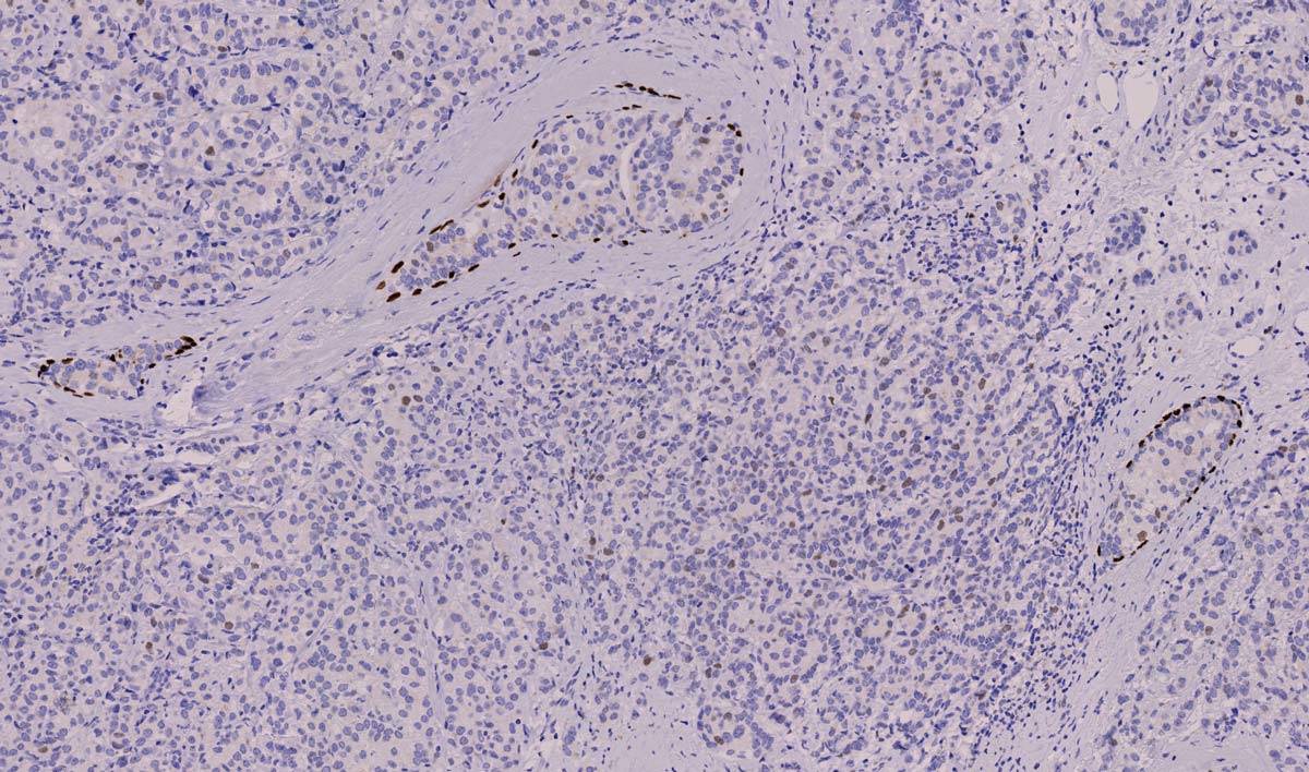
P63 Zoom
Analysis of serial sections with Virtual Multiplexing uses a patented method to apply and compare patterns across multiple slides. Here an IHC stained section for P63 is used to identify the stained cells.
02
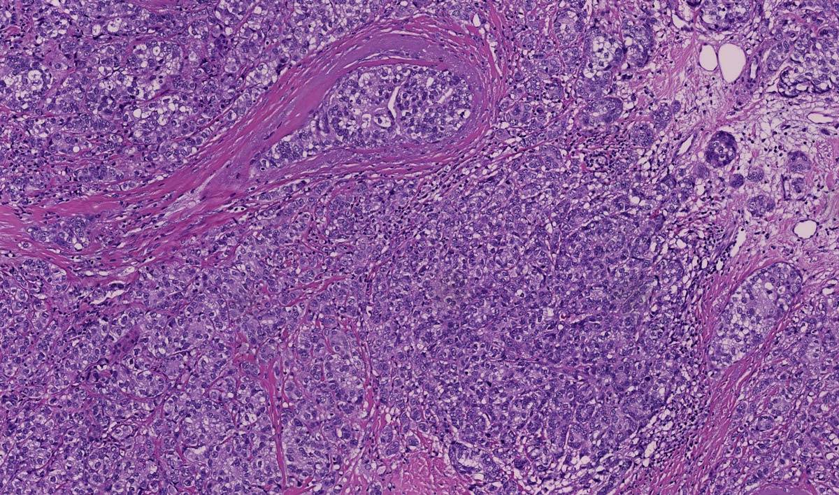
HE Zoom
H&E stained sections can be added to the analysis to verify morphology, assess regions of interest, identify metastatic regions and any regions can be applied across all serial sections
03
PCK Zoom
Cytokeratin stained sections help to quickly identify tumor versus stroma regions, and the identified regions can easily be applied to serial sections via virtual multiplexing of multiple tissue sections.
Improve your image analysis results with Visiopharm
Request demo
Tissue alignment has several advantages:
Could improve reproducibility, sensitivity, and specificity of automated quantitative biomarker assessments compared to manual reading
Enables tumor detection, using tumor markers such as cytokeratin, Melan-A, or other IHC) tumor markers. H&E stained tissue sections can also be useful for tumor detection
Enables robust, automated, and correct identification of invasive tumor cells, invasive tumor front, stroma, and pre-invasive tumor cells (incl. DCIS).
Allows co-localization studies of multiple biomarkers through virtual multiplexing
Quick and accurate alignment of any number of scanned tissue sections independently of image modality. Align IF with H&E or IHC brightfield to get the most our of your data.
Tissuealign™ is validated for in vitro diagnostic use (CE IVD) in Europe in combination with the CE IVD APPs from Visiopharm (ref. 1). All other applications are for Research Use Only.